People who go to their GP and are then suspected of having lung cancer are usually sent for an urgent chest X-ray. This is often performed within a few days.
The results of the X-ray may rule out lung cancer or they might show something suspicious.
Dr Gareth Roberts has many years experience of analysing chest X-rays and looking for signs that lung cancer may be present. As a General Respiratory Consultant in the NHS he is often called on to advise multidisciplinary teams made up of other health professionals.
In his private practice, Dr Roberts is able to offer expert advice on chest X rays that have already been obtained, or he can arrange a chest X-ray to investigate your symptoms.
He works closely with colleagues in other specialties to provide seamless care for patients as they are investigated and managed.
What happens next?
If something abnormal is found on your chest X-ray, it still does not confirm that you have lung cancer.
You will need further, more specialised tests to find out more.
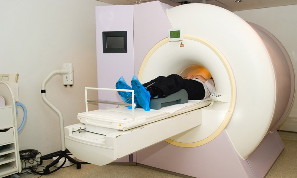
CT-scanning
This also uses X-ray technology but instead of taking one picture at one angle, it takes lots and produces a 3D image of the inside of your chest. It can reveal whether an abnormality on an X-ray is due to a tumour or scar tissue. If a tumour is present, the CT scan can show its exact position and how large it has become.
PET-CT scanning
This is a combination of a CT scan and a positron emission tomography scan (PET scan). Explained very simply, the PET scan follows the path of a radioactive substance that is injected into your vein. This tends to concentrate in tissue that is growing actively – such as a tumour. The PET scan detects where the radioactivity ends up, detecting a primary tumour and also showing if the cancer has spread through the lungs or beyond.
Flexible bronchoscopy
This involves looking at the inside of the airways with a flexible endoscope designed specially for the lungs.
Find out more information about flexible bronchoscopy here.
Fine needle aspiration biopsy
If all the tests performed indicate that you have a tumour within the lung, Dr Roberts will arrange for you to have a biopsy. This involves taking a small sample of the tumour so that the cells can be examined in a laboratory to find out exactly what type of lung cancer you have.
A lung biopsy is often performed in addition to a CT scan. The scan is used to reveal the exact position of the tumour so that a fine needle can be pushed through the skin and into the centre of the tumour. Biopsies are also performed using ultrasound guidance depending on the position of the tumour under investigation.
If your tumour is close to the airways, or within the airways, a biopsy can be taken during a flexible bronchoscopy.